Phase-Contrast Retinal Imaging

Phase contrast imaging reveals translucent structures by converting phase variations into detectable intensity differences. In the retina, this mechanism enables visualization of cells and microvascular structures that remain poorly contrasted in conventional reflectance imaging.

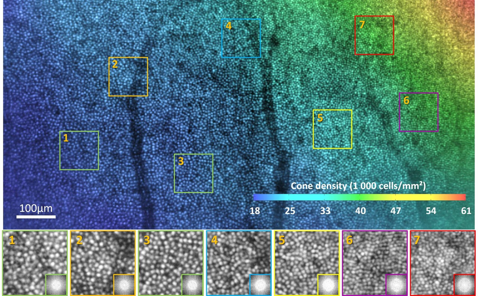
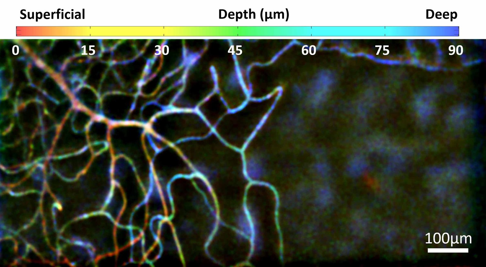
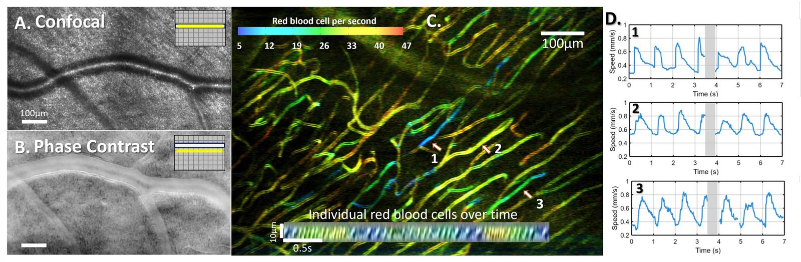
Our research develops phase-contrast retinal imaging through complementary optical architectures that tailor illumination and detection geometry to enhance contrast from translucent retinal structures. The Adaptive Optics Rolling Slit Ophthalmoscope (AO-RSO) combines line illumination with rolling shutter detection, enabling real-time contrast control and high-speed imaging over extended retinal areas. In parallel, we explore structured illumination strategies using digital micromirror devices (DMD), where patterned illumination combined with computational processing enables multimodal imaging from a single acquisition.

These approaches enable in vivo visualization of red blood cells, and vascular walls. By combining high temporal resolution with large-area imaging, they provide access to dynamic vascular processes such as blood flow, pulsatility, vasomotion, and stimulus-evoked vasodilation.

Illumination Geometry and Contrast Formation

We examine the physical mechanisms underlying retinal phase contrast and how illumination configuration and detection geometry determine image contrast. To do so, we develop computational models of light propagation in the eye, including a digital retinal twin that simulates scattering and image formation under different imaging conditions. These analyses clarify how translucent vascular structures can be visualized with enhanced contrast and guide the refinement of phase-contrast imaging for in vivo microvascular studies.

Adaptive Optics Rolling Slit Ophthalmoscopy (AO-RSO)

We developed the Adaptive Optics Rolling Slit Ophthalmoscope (AO-RSO) as a real-time phase-contrast retinal imaging system based on synchronized line illumination and rolling shutter detection. In this configuration, spatial filtering is performed optically at the camera level during acquisition, enabling efficient rejection of unwanted photons and flexible contrast control. By adjusting the relative position and width of the illumination line and detection slit, different imaging modes—including bright-field and phase-contrast—can be selected in real time within the same acquisition sequence. This architecture supports high-resolution, high-speed imaging over extended retinal areas and enables visualization of red blood cells, vascular walls, and dynamic microvascular processes in vivo. In parallel, we implement structured illumination strategies using digital micromirror devices (DMD), where controlled light patterns combined with computational reconstruction allow flexible modulation of image contrast from a single dataset.

Human retinal cone photoreceptors imaging using the AO-RSO (Adaptive Optics Rolling Slit Ophthalmoscope) system. Color codes the cone density.

Red blood cells imaging

In-vivo human retinal vascular plexuses at different depths (color-coded) using motion phase contrast imaging.

Vascular Dynamics

Phase-contrast retinal imaging enables direct visualization of red blood cells and microvascular walls in vivo. The high temporal resolution of adaptive optics imaging supports quantitative analysis of blood flow, velocity, pulsatility, and vasomotion across extended retinal areas. These capabilities allow dynamic vascular behavior to be measured under physiological conditions and during controlled visual stimulation. Together, they establish phase-contrast imaging as a powerful approach for studying retinal microcirculation at cellular resolution.

27/06/2025 in Neurovascular-Theme, Phase-Contrast-Theme, Publication

Revealing neurovascular coupling at a high spatial and temporal resolution in the living human retina

Pierre Senée, Léa Krafft, Inès Loukili, Daniela Castro Farias, Olivier Thouvenin, Michael Atlan, Michel Paques, Serge Meimon, and Pedro Mecê
Read More
23/12/2024 in Phase-Contrast-Theme, Publication

Adaptive Optics Rolling Slit Ophthalmoscope: Combining cellular-resolution, high-speed and large field-of-view in a multimodal retinal imager

Léa Krafft, Pierre Senee, Ana Alexandra Brad, Michael Atlan, Michel Paques, Olivier Thouvenin, Pedro Mecê, Serge Meimon
Read More
11/07/2022 in Phase-Contrast-Theme, Publication

Multimodal high-resolution retinal imaging using a camera-based DMD-integrated adaptive optics flood-illumination ophthalmoscope

Léa Krafft, Pierre Senée, Elena Gofas, Olivier Thouvenin, Michael Atlan, Michel Paques, Serge Meimon, and Pedro Mecê
Read More
06/12/2021 in Phase-Contrast-Theme, Publication

Design of a radial multi-offset detection pattern for in vivo phase contrast imaging of the inner retina in humans

Elena Gofas-Salas, Yuhua Rui, Pedro Mecê, Min Zhang, Valerie C. Snyder, Kari V. Vienola, Daniel M. W. Lee, José-Alain Sahel, Kate Grieve, and Ethan A. Rossi
Read More
29/10/2021 in Phase-Contrast-Theme, Publication

Partial-field illumination ophthalmoscope: improving the contrast of a camera-based retinal imager

Léa Krafft, Elena Gofas-Salas, Yann Lai-Tim, Michel Paques, Laurent Mugnier, Olivier Thouvenin, Pedro Mecê, and Serge Meimon
Read More
24/02/2021 in Phase-Contrast-Theme, Publication

Spatial-frequency-based image reconstruction to improve image contrast in multi-offset adaptive optics ophthalmoscopy

Pedro Mecê, Elena Gofas-Salas, Yuhua Rui, Min Zhang, José-Alain Sahel, and Ethan A. Rossi
Read More
06/07/2020 in Phase-Contrast-Theme, Publication

Optical Incoherence Tomography: a method to generate tomographic retinal cross-sections with non-interferometric adaptive optics ophthalmoscopes

Pedro Mecê, Elena Gofas-Salas, Michel Paques, Kate Grieve, and Serge Meimon
Read More
08/05/2019 in Phase-Contrast-Theme, Publication

Near infrared adaptive optics flood illumination retinal angiography

Elena Gofas-Salas, Pedro Mecê, Laurent Mugnier, Aurélie Montmerle Bonnefois, Cyril Petit, Kate Grieve, José Sahel, Michel Paques, and Serge Meimon
Read More

Pedro Mecê

PhD CNRS Researcher at Institut Langevin

PhD CNRS Researcher at Institut Langevin

Marcelina Sobczak

Postdoctoral Researcher

Marcelina Sobczak is a postdoctoral researcher specializing in retinal vascular dynamics and adaptive optics imaging. She leads research activities related to the characterization of neurovascular coupling and microvascular function within the group.
She obtained her PhD in Physics from Wroclaw University of Science and Technology, where her dissertation focused on the optical properties of the human cornea. She subsequently joined the laboratory of Stephen A. Burns at Indiana University, where she developed and optimized adaptive optics scanning laser ophthalmoscopy (AO-SLO) systems for the study of fast vascular dynamics and small vessel responses in vivo.
At CLARITY, she is responsible for advancing vascular functional imaging using the AO-RSO platform and for integrating new optical instrumentation to enhance the precision and temporal resolution of vascular measurements. She also contributes to the multimodal integration of interferometric (FFOCT) and phase-contrast imaging approaches to investigate the spatiotemporal relationship between neuronal activation and vascular responses at cellular resolution.

Clémence Baudet

PhD student

Clémence is a PhD candidate working on neurovascular functional imaging using adaptive optics phase-contrast imaging methods. Her research focuses on characterizing the spatiotemporal relationship between light-evoked neuronal responses and associated vascular dynamics in the living retina. She contributes to the implementation of new optical instrumentation on the AO-RSO platform to enable more precise and simultaneous measurements of neuronal activation and vascular responses.
She graduated from Institut d’Optique Graduate School before joining the group for her doctoral research.

Alejandra Superlano

PhD Student

Alejandra is a PhD candidate specializing in data-driven analysis for retinal imaging. She graduated with an Engineering Degree in Data Science and Healthcare from IMT Atlantique, France.
Her doctoral research focuses on the development of advanced AI–based image processing pipelines for retinal imaging and cellular-scale neurovascular coupling analysis. She is responsible for implementing computational strategies to enhance image quality, extract quantitative biomarkers, and support the analysis of spatiotemporal interactions between neuronal and vascular signals.

Andrès Cortes

Master 2 student

Get In Touch

Interested or need more details? Leave us a message and we’ll get back to you shortly.

    Support

    Logo
    Logo
    Logo
    Logo
    Logo