Neurovascular Functional Imaging

The retina is the only part of the central nervous system that can be imaged non-invasively at cellular resolution. This unique accessibility enables simultaneous measurement of light-evoked neuronal responses and associated microvascular dynamics in vivo.

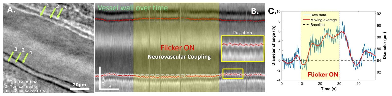
Building on interferometric and phase-contrast imaging approaches developed in the group, we investigate how neuronal activation and vascular responses can be measured and quantified concurrently. OCT-based interferometric imaging detects photoreceptor activity, while phase-contrast imaging reveals vascular structure and blood cell motion with high temporal resolution. By integrating these complementary measurements, we analyze the spatio-temporal relationships between neuronal activity and hemodynamic regulation.

This work aims to establish quantitative frameworks for characterizing neurovascular interactions in the human retina and to understand how cellular-scale neuronal responses are coupled to dynamic vascular behavior in health and disease.

Characterization of Vascular Response Dynamics

High-speed phase-contrast AO-RSO imaging enables continuous measurement of arterial diameter dynamics in vivo, both at rest and during visual stimulation. This approach separates cardiac pulsation, spontaneous vasomotion, and stimulus-evoked dilation within the same acquisition. The resulting measurements provide quantitative characterization of the temporal structure and amplitude of neurovascular responses in the human retina.

Stimulus- and Vessel-Dependent Vascular Responses

We examine how retinal vascular responses vary with stimulation parameters and vessel type. By analyzing variations in response amplitude and temporal dynamics across conditions and vascular segments, we characterize structured patterns of reactivity within the retinal microcirculation. These studies contribute to a quantitative framework for understanding neurovascular regulation in vivo.

27/06/2025 in Neurovascular-Theme, Phase-Contrast-Theme, Publication

Revealing neurovascular coupling at a high spatial and temporal resolution in the living human retina

Pierre Senée, Léa Krafft, Inès Loukili, Daniela Castro Farias, Olivier Thouvenin, Michael Atlan, Michel Paques, Serge Meimon, and Pedro Mecê
Read More

Pedro Mecê

PhD CNRS Researcher at Institut Langevin

Principal Investigator, CLARITY Research Group
Pedro Mecê is a CNRS Research Scientist at Institut Langevin (CNRS/ESPCI Paris – PSL), and co-leader of the CLARITY Research Group. He leads research on in vivo retinal imaging using OCT-based interferometric and phase-contrast optical methods. His work lies at the interface between optical physics and biomedical imaging, with the ambition of transforming retinal imaging into a tool for probing neuronal and vascular function at cellular resolution.

He completed a CIFRE PhD in collaboration with ONERA and Quantel Medical, focusing on adaptive optics–assisted retinal laser surgery with micrometric precision. He subsequently joined Institut Langevin as a postdoctoral researcher, where he pioneered the implementation of time-domain full-field OCT for in vivo human retinal imaging. In 2022, he was appointed CNRS Research Scientist to develop new optical architectures, wavefront engineering strategies, and computational imaging approaches aimed at extracting structural and functional biomarkers from the living human retina.

His research is driven by the unique accessibility of the retina as the only optically transparent window into the central nervous system, enabling direct investigation of neuronal and vascular networks in vivo at cellular scale.

His work has been recognized by several scientific awards, including thesis prizes from the French Microscopy Society (Sfµ) and the French Society for Biological and Medical Engineering (SFGBM), as well as international distinctions from OPTICA, SPIE and ARVO. He is the recipient of the ANR JCJC BRAINS project (2023-2027) and currently leads the ERC MIRACLE-AD (2026-2031). He is co-founder of the start-up SharpEye and an active member of the Paris Eye Imaging Group, which brings together physicists and clinicians at the Quinze-Vingts National Eye Hospital.

Download full CV

Olivier Thouvenin

PhD, Hab., Assistant Professor at ESPCI

Olivier Thouvenin is Associate Professor of Optics at ESPCI Paris – PSL and co-leader of the CLARITY Research Group. His research focuses on label-free optical microscopy, quantitative imaging, and biophysical signal analysis. He develops advanced full-field optical methods and data-processing strategies to investigate dynamic biological processes without exogenous contrast agents.
He obtained a Master’s degree in Biophysics from ENS Lyon and Université Paris 7 in 2014 and completed his PhD in Optical Microscopy in 2017, during which he co-invented Dynamic Full-Field OCT (D-FFOCT). After postdoctoral work studying cerebrospinal fluid mechanics and its role in zebrafish scoliosis models, he joined ESPCI Paris as Assistant Professor, where he now leads a research group dedicated to label-free imaging and quantitative biophysics.
His contributions have been recognized internationally. In 2018, Nature identified him as one of eleven international rising stars. He received the Prix Edouard Branly in 2023 and the Prix des Innovateurs from Région Île-de-France in 2024. In 2024, he co-founded Lutèce Dynamics, a company dedicated to the dissemination of Dynamic FFOCT technologies for biology and pharmacology.

Inès Loukili

Postdoctoral Researcher

Inès is currently a postdoctoral researcher (former PhD student) in our group. Her work focuses on the theoretical and computational modeling of interferometric retinal imaging for both structural and functional applications. She develops analytical frameworks and numerical simulations to describe how spatial and temporal coherence influence image formation and performance in full-field and line-field OCT systems. Her contributions provide the quantitative foundations that guide the design and optimization of coherence-engineered approaches for in vivo retinal imaging.
She completed her engineering degree at Institut d’Optique Graduate School, including a double-degree program with KTH Royal Institute of Technology, before pursuing her PhD within the group.

Marcelina Sobczak

Postdoctoral Researcher

Marcelina Sobczak is a postdoctoral researcher specializing in retinal vascular dynamics and adaptive optics imaging. She leads research activities related to the characterization of neurovascular coupling and microvascular function within the group.
She obtained her PhD in Physics from Wroclaw University of Science and Technology, where her dissertation focused on the optical properties of the human cornea. She subsequently joined the laboratory of Stephen A. Burns at Indiana University, where she developed and optimized adaptive optics scanning laser ophthalmoscopy (AO-SLO) systems for the study of fast vascular dynamics and small vessel responses in vivo.
At CLARITY, she is responsible for advancing vascular functional imaging using the AO-RSO platform and for integrating new optical instrumentation to enhance the precision and temporal resolution of vascular measurements. She also contributes to the multimodal integration of interferometric (FFOCT) and phase-contrast imaging approaches to investigate the spatiotemporal relationship between neuronal activation and vascular responses at cellular resolution.

Anita Mouttou

Research Engineer

Anita Mouttou is a research engineer at CLARITY specializing in the design and integration of advanced interferometric imaging systems. She is responsible for the development of a line spectral-domain OCT platform dedicated to ultra-sensitive measurements of retinal dynamics. Her work focuses on optimizing interferometric sensitivity, system stability, and high-speed acquisition to detect subtle structural and functional variations in the living retina.

She graduated from Institut d’Optique Graduate School and completed her PhD developing resonant dielectric multilayer architectures for sensitivity enhancement in TIRF microscopy, enabling high-precision single-molecule imaging in living cells.

Oliver Martinache

PhD Student

Olivier is a PhD candidate working on structural and functional retinal imaging using interferometric modalities. His research explores retinal dynamics through digital holography and time-domain full-field OCT, with a focus on quantifying optoretinographic responses, cellular motion, and retinal blood flow. Within the group, he leads the experimental implementation of optoretinography in the TD-FFOCT platform, including amplitude-based ORG strategies, spectral shaping, and system optimization for in vivo measurements.
He graduated from Institut d’Optique Graduate School before joining the group for his doctoral research.

Clémence Baudet

PhD student

Clémence is a PhD candidate working on neurovascular functional imaging using adaptive optics phase-contrast imaging methods. Her research focuses on characterizing the spatiotemporal relationship between light-evoked neuronal responses and associated vascular dynamics in the living retina. She contributes to the implementation of new optical instrumentation on the AO-RSO platform to enable more precise and simultaneous measurements of neuronal activation and vascular responses.
She graduated from Institut d’Optique Graduate School before joining the group for her doctoral research.

Alejandra Superlano

PhD Student

Alejandra is a PhD candidate specializing in data-driven analysis for retinal imaging. She graduated with an Engineering Degree in Data Science and Healthcare from IMT Atlantique, France.
Her doctoral research focuses on the development of advanced AI–based image processing pipelines for retinal imaging and cellular-scale neurovascular coupling analysis. She is responsible for implementing computational strategies to enhance image quality, extract quantitative biomarkers, and support the analysis of spatiotemporal interactions between neuronal and vascular signals.

Elise Couturier

Master 2 student

Elise is a Master’s student intern. She is currently pursuing an optical engineering diploma at Institut d’Optique Graduate School, specializing in Biomedical Engineering, and has completed part of her training in Madrid.
She works on spatial coherence engineering strategies aimed at enhancing resolution and sensitivity in full-field interferometric imaging.

Andrès Cortes

Master 2 student

Get In Touch

Interested or need more details? Leave us a message and we’ll get back to you shortly.

    Support

    Logo
    Logo
    Logo
    Logo
    Logo