Researchers from the CLARITY Group at Institut Langevin (ESPCI Paris, CNRS, PSL University), in collaboration with Quinze-Vingts National Eye Hospital and ONERA, have developed a breakthrough optical imaging method that enables direct observation of retinal neurovascular coupling at cellular resolution in the living human eye.
Neurovascular coupling is a fundamental physiological process through which neural activity locally regulates blood flow to ensure adequate delivery of oxygen and nutrients. This mechanism underlies functional brain imaging techniques such as fMRI, yet it had never previously been measured at the cellular scale in humans.
This advance was made possible by imaging the retina, the only part of the central nervous system that is optically accessible in vivo. As a direct extension of the brain, the retina offers a unique, non-invasive window for investigating neurovascular processes at microscopic resolution.
To achieve this, the researchers developed the Adaptive Optics Rolling Slit Ophthalmoscope (AO-RSO). This novel system synchronises line illumination with the rolling shutter of a two-dimensional scientific camera. By introducing a controlled spatial offset between illumination and detection, the instrument generates label-free phase-contrast images of the retina, enabling measurements of vessel diameter changes with 100-nanometre spatial precision and temporal resolution down to 10 milliseconds.
Using this technique, the team isolated vascular responses specifically induced by light-evoked neural activity, distinct from physiological fluctuations such as vasomotion or cardiac pulsation. In healthy volunteers, the vascular response was shown to follow a triphasic temporal profile, with characteristics that depend on vessel diameter, stimulus duration, and vascular location.
By opening a non-invasive, cellular-resolution window onto neurovascular function, this work holds strong potential for the early detection of neurovascular and neurodegenerative diseases, including Alzheimer’s disease and diabetic retinopathy. The study was supported by the ANR BRAINS project and IHU FOReSIGHT, and was published in Science Advances.
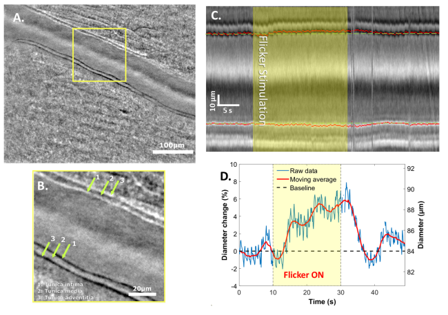
(A) Phase contrast image of a retinal artery acquired with the AO-RSO system.
(B) Magnified view showing the structure of the vessel wall.
(C) Kymograph of the artery during flicker light stimulation (highlighted by the yellow rectangle). The green dashed line indicates the baseline diameter.
(D) Time course of arterial diameter in response to flicker stimulation.